Cross-Modality Transformations in Biological Microscopy
Seeing Fluorescence Without the Fluorophores
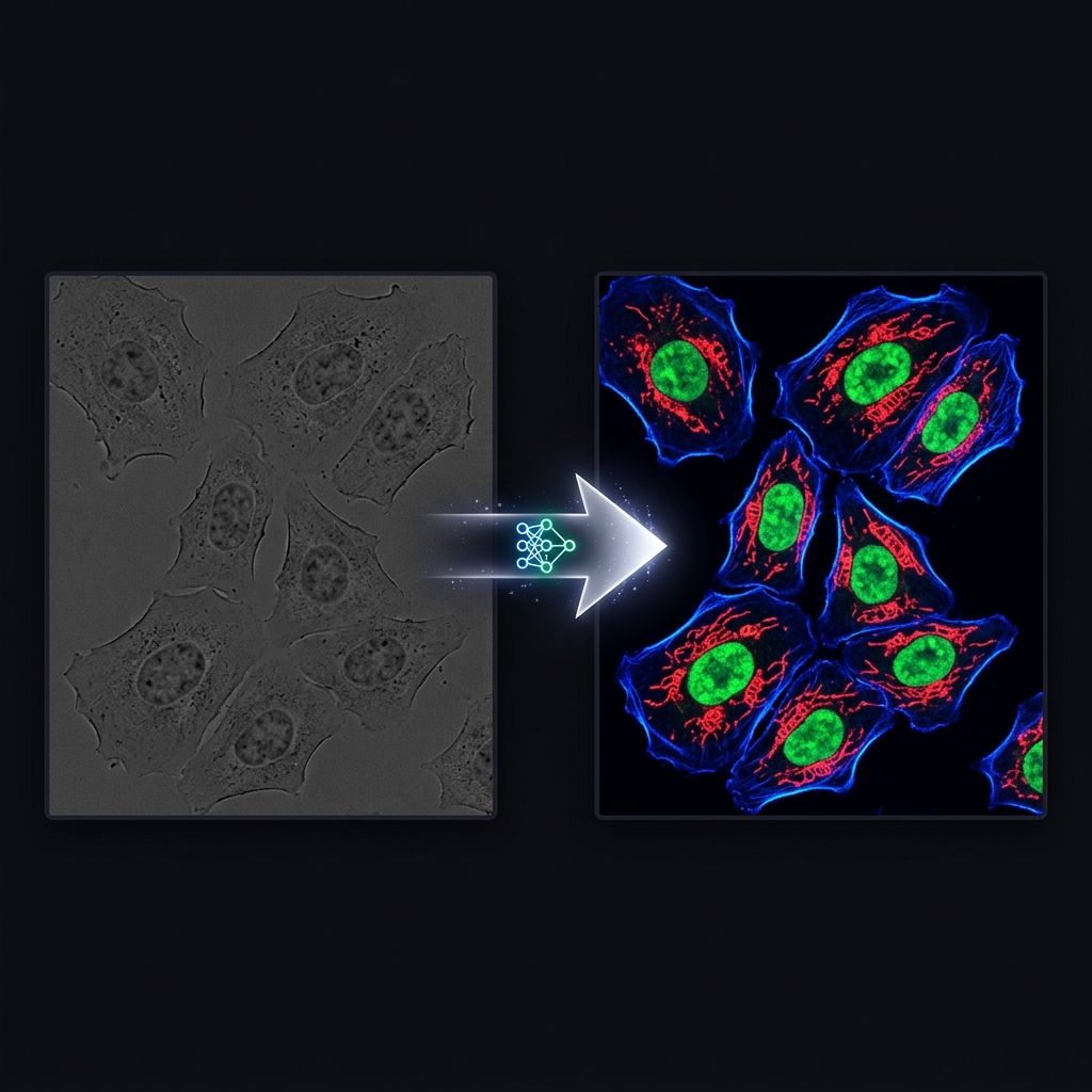
Biological imaging often requires researchers to choose between cellular health and image contrast. Brightfield microscopy is non-invasive but offers low contrast. Fluorescence microscopy highlights specific structures beautifully but requires chemical stains that are phototoxic and alter cellular behavior.
In our 2024 publication in Advanced Photonics, Cross-modality transformations in biological microscopy enabled by deep learning, we explored how to get the best of both worlds using generative AI.
In Silico Staining
Using Generative Adversarial Networks (GANs) and diffusion models, we trained architectures to perfectly translate simple, non-invasive brightfield images into high-contrast "virtual" fluorescence images.
Because brightfield images contain latent phase information about cellular structures (like the nucleus or mitochondria), the deep learning model can mathematically infer where a fluorescent dye would bind, generating a synthetic fluorescent image with immense accuracy.
Key Advances from the Research:
- Zero Phototoxicity: Researchers can observe complex cellular structures over long time-lapses without exposing the cells to harmful UV light or chemical dyes.
- Multiplexing: A single brightfield scan can be synthetically transformed into multiple different fluorescent channels simultaneously.
- High Fidelity: The AI-generated images exhibit structural correlations exceeding 95% when compared to true physical ground-truth fluorescence.
Revolutionizing High-Content Screening
For IFLAI's clients in pharma and biotech, this capability drastically reduces the cost and complexity of phenotypic screening assays. By leveraging "in silico staining," laboratories can accelerate high-throughput drug screening workflows while maintaining the absolute physical integrity of their live-cell cultures.