Giving Microscopes a Brain: The Rise of Agentic AI in the Lab
Most people still imagine AI as something that happens after the experiment.
You acquire the data. You export the images. You run the model. The model detects cells, segments particles, classifies phenotypes, scores morphology, or generates a report. In that version of the story, the instrument does the experiment and the AI analyzes the result.
That view is already becoming outdated.
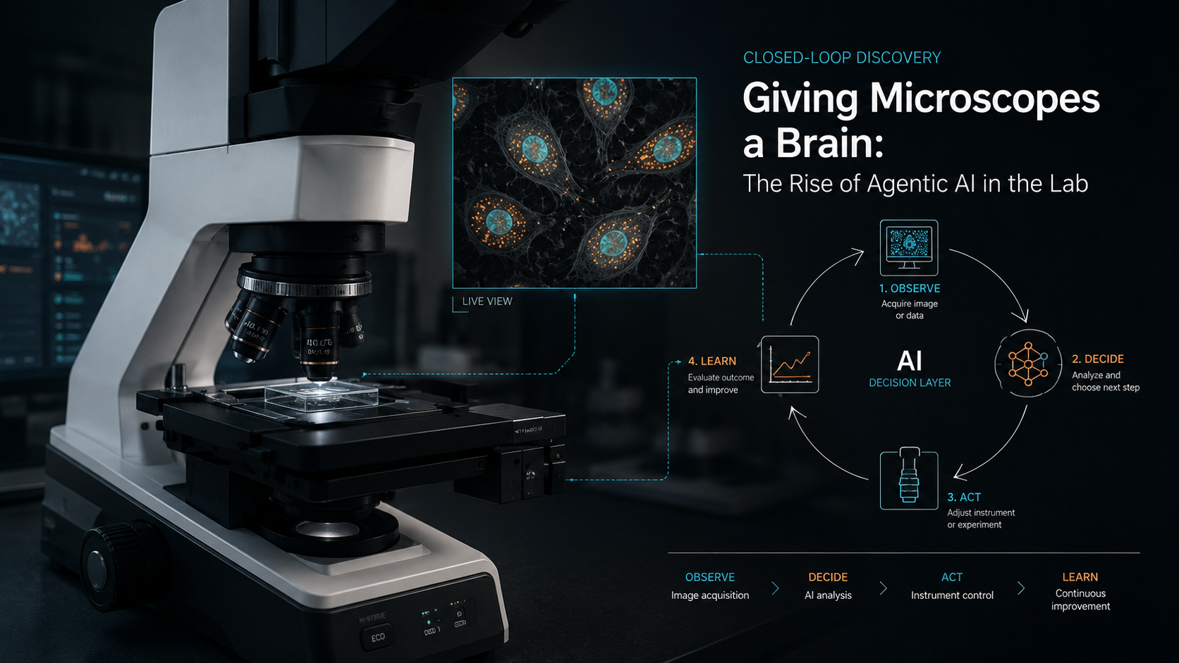
The more interesting future is not AI as a passive analysis layer. It is AI as an experimental interface: a system that can observe what is happening, decide what is worth measuring next, adjust the instrument, and close the loop between data collection and learning.
In microscopy, that means a model does not just analyze an image after acquisition. It can decide where the microscope should move, whether the focus is good enough, which cells are interesting, whether more resolution is needed, whether a region should be revisited, whether the current field of view is uninformative, or whether the experiment has already collected enough evidence to answer the question.
That is the real meaning of giving microscopes a brain. Not making them magical. Not replacing scientists. But connecting perception, decision-making, and instrument control into a closed loop.
From Analysis to Action
The first wave of AI in scientific imaging was mostly about interpretation. Models became better at identifying objects, segmenting cells, denoising images, extracting features, and building representations from complex data. That was already transformative, because microscopy produces far more visual information than humans can inspect manually.
But interpretation is only one part of the experiment.
A human microscopist does not simply stare at a screen. They make decisions constantly. They move the stage, adjust the focus, change exposure, avoid dead regions, zoom into interesting structures, repeat uncertain measurements, and stop when the experiment has produced enough useful information. A good experimentalist is not just a detector. They are an active controller of the measurement process.
That is the gap agentic AI begins to close.
An agentic laboratory system is not merely asked, “What is in this image?” It is asked, “Given what we have seen so far, what should we do next?” In that small change, AI moves from classification to experimentation.
This shift is already visible in the broader self-driving laboratory field, where autonomous platforms combine robotics, machine learning, experimental planning, and data analysis into closed discovery loops. Recent reviews describe self-driving laboratories as systems that automate experimental workflows and use autonomous planning to accelerate chemistry and materials discovery [1].
Microscopy is a particularly natural place for this transition, because the instrument already has controllable degrees of freedom. The stage can move. The objective can change. The focus can be adjusted. Illumination can be tuned. Exposure can be modified. Acquisition can be sparse, dense, fast, slow, low-resolution, high-resolution, fixed, adaptive, destructive, or gentle.
The question is no longer whether AI can analyze microscopy data. The question is whether AI can help decide how the microscopy data should be collected in the first place.
The Closed-Loop Microscope
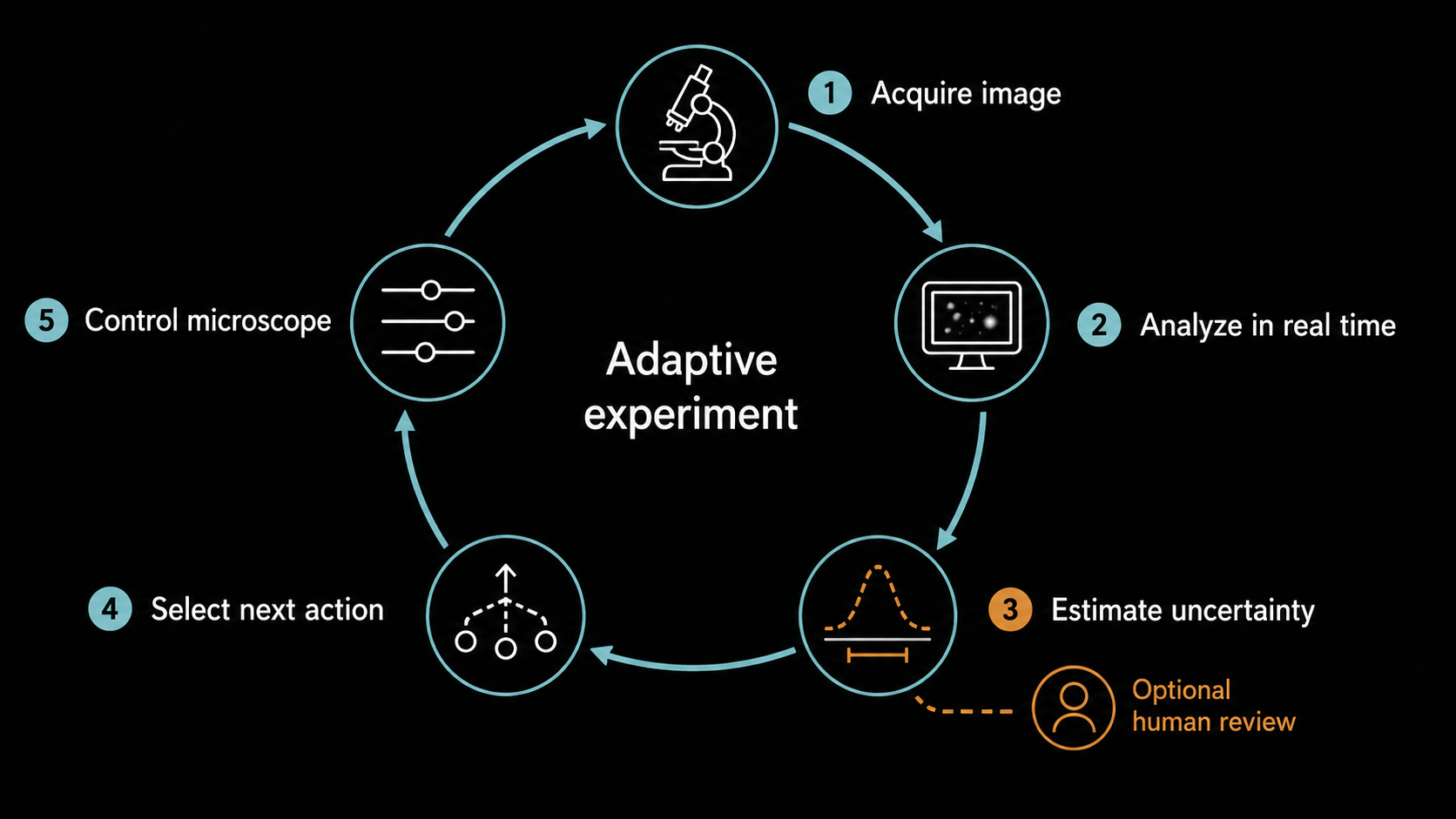
A closed-loop microscope has four basic parts. It observes the sample. It interprets what it sees. It decides what action would be most useful next. Then it executes that action through the instrument.
Observe. Interpret. Decide. Act. Learn.
That loop can be simple. For example, the system may detect that a cell has drifted out of frame and move the stage to keep it centered. Closed-loop imaging strategies have already been used to automatically compensate for cell movement by tracking position and adjusting experimental hardware in real time [2].
But the same principle can become much more powerful. A microscope can prioritize rare events, reduce unnecessary imaging, avoid phototoxicity, focus acquisition on uncertain regions, or actively search for structures that are likely to teach the model something new. Smart microscopy is already moving in this direction by combining real-time image analysis with adaptive acquisition, and recent work has extended this idea toward outcome-driven microscopy, where imaging and perturbation are used to steer biological processes toward predefined outcomes [3].
In materials science, autonomous microscopy has developed even further. Active-learning approaches in scanning probe microscopy have been used to guide nanoscale exploration, choose where to measure next, and discover structure-property relationships more efficiently than static acquisition strategies. The SEEK framework, for example, incorporates expert knowledge into autonomous scanning probe microscopy workflows so that the system explores samples in a more targeted way [4].
This is the core idea: the microscope should not waste time measuring blindly.
If the goal is to understand a heterogeneous sample, most fields of view are not equally valuable. Some are redundant. Some are artefactual. Some are low quality. Some contain rare events. Some sit exactly at the boundary where the model is uncertain. A passive pipeline treats all acquired data as fixed. An agentic microscope can ask which measurement would be most useful next.
That is where active learning becomes more than a machine-learning technique. It becomes an experimental strategy.
Why Tool Protocols Matter
For this future to work, AI systems need a reliable way to communicate with instruments, software, databases, and analysis pipelines.
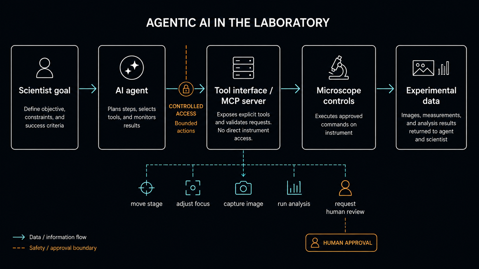
This is where tool-use standards such as the Model Context Protocol (MCP) are interesting. MCP was introduced by Anthropic in 2024 as an open standard for building secure two-way connections between AI-powered tools and external data sources. Its specification describes a structure in which servers can expose resources, prompts, and tools; tools are functions the AI model can execute, while resources provide contextual data such as files, schemas, or application-specific information [5, 8].
In ordinary software settings, that might mean connecting an AI assistant to GitHub, Slack, a database, or a file system. In the laboratory, the same architectural idea becomes much more interesting.
A microscope could expose tool calls such as:
move_stage(x, y)set_focus(z)change_objective(magnification)capture_image(channel, exposure)run_segmentation(image_id)score_cell_state(region_id)select_next_region(strategy)pause_for_human_review(reason)
The model would not need to “be” the microscope. It would operate through a controlled interface, where each action is explicit, logged, constrained, and reversible where possible. That distinction matters. Agentic AI in the lab should not mean giving a language model unrestricted control over expensive equipment. It means building a structured control layer where AI can propose or execute bounded actions under clear rules.
This is what makes the concept practical.
The future laboratory agent is not a chatbot bolted onto a microscope. It is an orchestration layer sitting between the scientist, the instrument, the data, and the learning system. It can translate a high-level goal into a sequence of experimental actions, but those actions must pass through instrument-specific constraints, safety rules, permission boundaries, and audit logs.
In that sense, MCP and related tool-use protocols are not the whole solution. They are part of the plumbing that makes the solution possible.
The Scientist Moves Up the Stack
The common fear around autonomous laboratories is that they remove the scientist from the experiment. That is the wrong way to think about it.
A good agentic lab system should remove repetitive control burden, not scientific judgement. It should not replace the scientist’s role in deciding what matters, what hypothesis is worth testing, or whether the result makes sense. It should instead move the scientist away from low-level operational decisions and toward higher-level experimental strategy.
Instead of manually inspecting hundreds of fields of view, the scientist defines the objective:
- Find rare phenotypes.
- Track these cells over time.
- Maximize information while minimizing phototoxicity.
- Explore this material surface until uncertainty falls below a threshold.
- Ask for review when the system sees something outside its expected distribution.
The AI then handles the tedious middle layer: continuous monitoring, prioritization, acquisition decisions, quality checks, and adaptation.
This changes the rhythm of experimentation. Instead of running a fixed protocol and discovering afterwards that half the images were uninformative, the experiment can adjust while it is still running. Instead of collecting massive datasets first and asking questions later, the system can collect data because it is informative for the question.
That is a much more efficient way to use both instruments and human attention.
Why This Matters for Data-Efficient AI
Agentic microscopy connects directly to data efficiency. If AI only analyzes data after acquisition, then the dataset has already been shaped by manual choices, fixed protocols, and whatever happened during the run. The model can learn from the dataset, but it cannot influence how the dataset is created.
Once AI enters the acquisition loop, that changes.
The system can choose examples that reduce uncertainty. It can avoid collecting thousands of redundant images. It can request expert labels only for regions where labels are likely to matter. It can adapt acquisition when the sample behaves unexpectedly. It can discover that the model is failing under a specific condition and collect targeted data to repair that weakness.
This is the difference between data collection and data strategy.
In a passive workflow, more data often means more storage, more annotation, more compute, and more cleanup. In an active workflow, data become more intentional. Each measurement has a reason to exist.
This is especially important in scientific imaging, where data are not free. Imaging time costs money. Labels require expertise. Live-cell imaging can damage samples. High-resolution acquisition may be slow. Large experiments create bottlenecks in storage, compute, and quality control. A system that collects less data but more useful data can be better than one that collects everything.
That is why agentic AI in the lab is not just an automation story. It is a data-efficiency story.
The Hard Part is Not Making the Agent Act
It is tempting to describe agentic AI as if the main breakthrough is simply allowing the model to use tools. But in laboratories, the hard part is not making software take an action. The hard part is making sure it takes the right action, for the right reason, inside the right boundary.
A lab agent needs uncertainty estimates. It needs quality control. It needs a model of what the instrument can and cannot do. It needs to distinguish scientific signal from technical artefact. It needs to know when to stop, when to ask for help, and when its own predictions are unreliable.
It also needs security and governance. Tool-connected AI systems introduce new risks, including prompt injection, tool poisoning, weak isolation, and unintended access to external systems. Recent MCP security analyses have highlighted tool poisoning as a serious vulnerability, where malicious instructions can be embedded in tool metadata rather than in the user’s visible prompt [6].
In a lab, these risks are not abstract. An unsafe agent should not be able to damage equipment, overwrite data, run uncontrolled experiments, or silently change acquisition parameters. The more physical the AI becomes, the more important it is that autonomy is bounded.
The right design is not maximum autonomy. It is calibrated autonomy.
Some actions can be fully automatic. Some should require confirmation. Some should be impossible. A system may be allowed to move the stage within a predefined region, but not change laser power beyond a safe range. It may select fields of view, but not discard raw data. It may recommend a perturbation, but require human approval before applying it. It may continue acquisition overnight, but pause if the sample moves outside the expected distribution.
This is how laboratory AI should evolve: not as an uncontrolled agent, but as a capable experimental operator with clearly defined authority.
From Smart Microscopes to Scientific Co-pilots
The most exciting version of agentic microscopy is not a fully automated black box. It is a scientific co-pilot that understands enough about the instrument, the sample, and the objective to make experimentation faster and more adaptive.
The scientist still defines the question. The system helps execute the search. This is a very different relationship with laboratory equipment. The microscope becomes less like a camera and more like an active experimental system. It does not only capture the sample. It participates in the measurement strategy.
That is where modern AI becomes most useful in the physical sciences and life sciences: not as a detached model that produces outputs after the fact, but as a loop that connects perception, decision-making, and action.
Giving Microscopes a Brain
The next step in AI for scientific imaging is not only better segmentation, better classification, or better representation learning. Those still matter, but they are no longer the whole story.
The next step is closing the loop.
AI systems will not only analyze what the microscope has already seen. They will help decide what the microscope should look at next. They will use uncertainty, prior knowledge, active learning, and real-time feedback to make experiments more adaptive. They will connect models to instruments through structured tool interfaces. They will turn data acquisition from a fixed protocol into an intelligent process.
At IFLAI, this is the direction we believe matters: data-efficient, physics-informed AI that does not stop at analysis, but connects to the experimental workflow itself.
The future microscope will not just see more. It will know where to look.
References
- [1] American Chemical Society Publications, "Self-Driving Laboratories for Chemistry and Materials Science"
- [2] PMC, "Closed-loop real-time imaging enables fully automated cell tracking and adaptive acquisition"
- [3] Nature, "Closed-loop optogenetic control of cell biology enables outcome-driven acquisition"
- [4] RSC Publishing, "Scientific exploration with expert knowledge (SEEK) in autonomous scanning probe microscopy"
- [5] Anthropic, "Introducing the Model Context Protocol"
- [6] arXiv, "Model Context Protocol Threat Modeling and Analyzing Vulnerabilities to Prompt Injection with Tool Poisoning"
- [7] RSC Publishing, "Autonomous laboratories for accelerated materials discovery"
- [8] ModelContextProtocol.io, "Specification"