Kommentar på Clines Scientifics inledda samarbete med IFLAI gällande utveckling av AI-mjukvara för CellRace-projektet
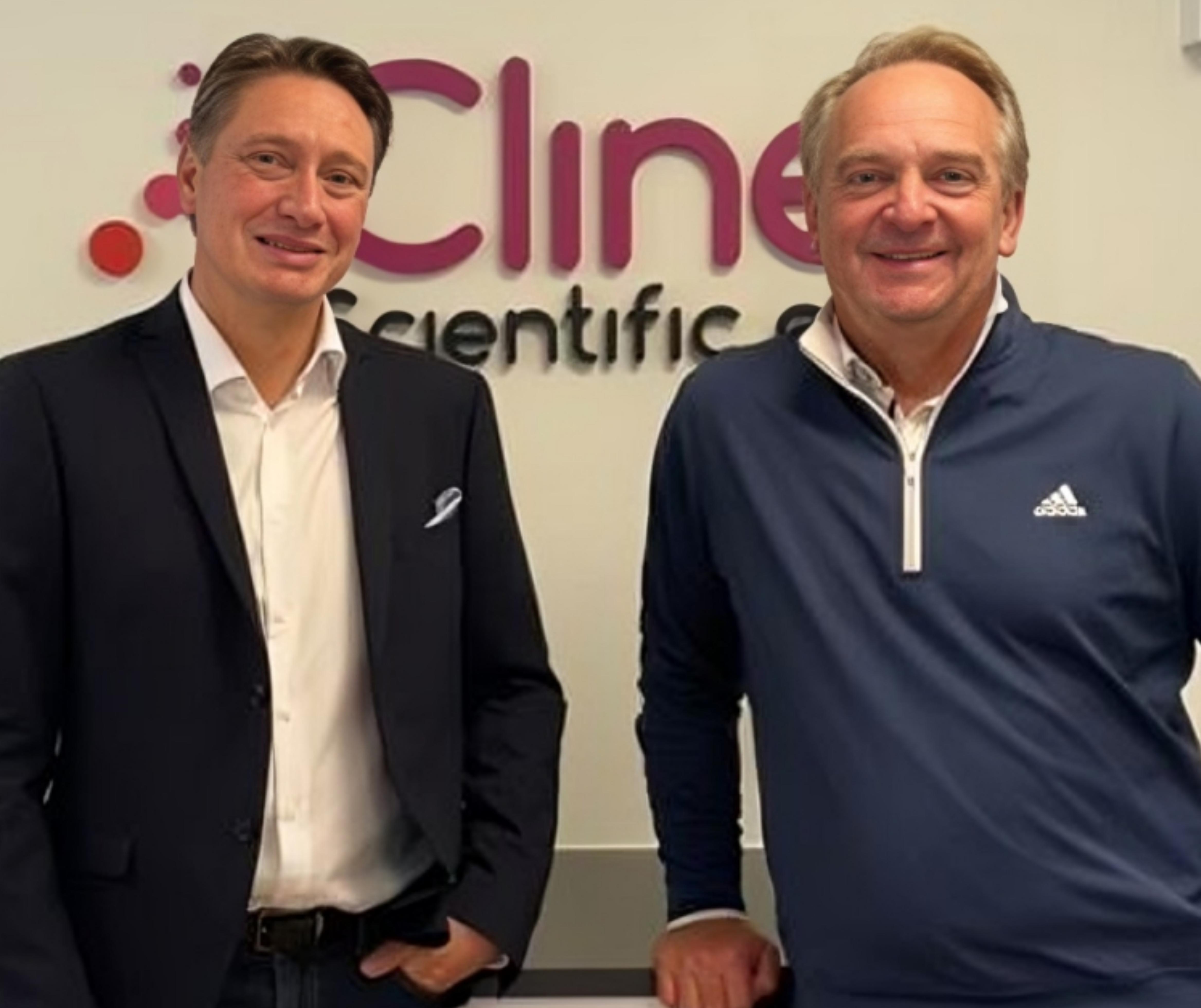
Home Publications Investors News Get Started Kommentar på Clines Scientifics inledda samarbete med IFLAI gällande utveckling av AI-mjukvara för CellRace-projektet Cline Scientific AB (”Cline” eller ”bolaget”) tillkännagav idag den 7:e oktober att bolaget initierat ett samarbete med mjukvaruföretaget IFLAI som är specialiserade på AI-lösningar inom Life Science för att utveckla en skräddarsydd AI-baserad mjukvara för cancerdiagnostikprojektet CellRace som bygger på just avancerade algoritmer i syfte att bedöma och förutse risken för metastaser. I ett första steg ska Cline tillsammans med IFLAI samarbeta för att skapa en skräddarsydd lösning för den automatiserade analysen av den data som samlats in i CellRACE-projektet för att underlätta vidare-utveckling. Detta delmoment kommer att slutföras under Q4-22, därefter kommer Cline att testa program-varan och samla in data från en rad olika tumörcelltyper. I ett efterföljande projekt kommer denna data att användas för att förbättra systemet eftersom det kan ”lära sig” ett brett spektrum av cancerfenotyper, och sedan i slutändan användas som en del av en integrerad algoritm och mätsystem för att bestämma cancermetastatisk potential från ett patientprov. Vår syn på Clines nya samarbetet är att det är naturligt och viktigt strategiskt steg då utvecklandet av avancerade AI-lösningar är en komplex uppgift som kräver många olika kompetenser och specialiteter. Genom att Clines djupgående kunskap inom biomedicin och bioteknologi kombineras med IFLAI:s expertis inom artificiell intelligens med fokus på Life Science innovationer ser vi att intressanta nyheter och data kan presenteras framgent gällande CellRACE-projektet. Professorerna Mattias Goksör och Giovanni Volpe som representerar IFLAI har sammantaget publicerat cirka 100 forskningsartiklar med över 14 000 citeringar enligt forskningsdatabasen Google Scholar. Read more Recent Posts November 25, 2023 IFLAI’s Chief AI Engineer spearheads AI dialogue at Future Impact Summit 2023 September 18, 2023 Giovanni Volpe awarded the Faculty of Science’s 2023 Research Award April 5, 2023 IFLAI participates in GAIA Conference 2024, sharing the stage with global AI leaders February 20, 2023 Giovanni Volpe Shares Machine Learning Expertise at ICFO January 24, 2023 IFLAI has been awarded the prestigious ERC Proof of Concept Grant Publications Investors News Contact Us AddressHasselvägen 85, Mölnlycke Emailcontact@iflai.com Get Connected
Cline starts collaboration with IFLAI in development of AI software for CellRACE project
Home Publications Investors News Get Started Cline starts collaboration with IFLAI in development of AI software for CellRACE project Cline Scientific announces that it initiates a project with software company IFLAI to develop custom AI-based software for the cancer diagnostic project, CellRACE. Since earlier in 2022, researchers at the University of Gothenburg and Chalmers University of Technology have been collaborating with Cline in exploring whether it is possible to apply algorithms centered around deep learning (a type of machine learning/artificial intelligence) to track and analyze tumor cell behavior and features automatically and objectively. The initial work showed that such a method can be used effectively as a basis for an automated software solution. Now, Cline takes the next step and outsources the development of an AI software solution to IFLAI, a company with roots in the academic community and documented know-how in this field. Cline and IFLAI have agreed on a first-step project to create a tailored solution to the automated analysis of data gathered in the CellRACE project to aid in CellRACE’s further development. This project will be completed in Q4 of 2022. After this, the Cline team will test the software and gather data on a range of tumor cell types. In a following project, this data will be used to enhance the system as it can “learn” a wide range of cancer phenotypes and then ultimately be used as part of an integrated algorithm and measurement system to determine cancer metastatic potential from a patient sample. “We are extremely proud that Cline Scientific have chosen our AI-software for its image analysis work. Cline Scientific is an ideal client for us at IFLAI: a well-established company trying to bring a groundbreaking new technology into the forefront of the industry, limited mainly by the common bottleneck of data analysis. With our AI-based tools, we can push the limits in terms of speed, accuracy, reproducibility and scalability of CellRACE analysis.” – Professor Mattias Goksör, CEO of IFLAI. About CellRACECline is developing CellRACE, a cell-based cancer diagnostic product that can assess the risk of metastasis. Using the Cline gradient surfaces combined with advanced analytics, CellRACE aims to predict the probability of metastasis in an accurate manner not possible today as well as in a much earlier stage so the patient can start relevant treatment. The product has potential to be used both in breast cancer diagnosis as well as in other forms of metastasizing cancer. The analysis method forms part of the overall CellRACE project and its pathway to becoming a marketed product. Once the analytical method and prediction algorithm has been completed, the next milestone in the CellRACE development journey will consist of conducting a pilot study. Following this, continued product development and final product design will be completed before performing the necessary regulatory approval steps, such as a clinical performance evaluation. Read more Recent Posts November 25, 2023 IFLAI’s Chief AI Engineer spearheads AI dialogue at Future Impact Summit 2023 September 18, 2023 Giovanni Volpe awarded the Faculty of Science’s 2023 Research Award April 5, 2023 IFLAI participates in GAIA Conference 2024, sharing the stage with global AI leaders February 20, 2023 Giovanni Volpe Shares Machine Learning Expertise at ICFO January 24, 2023 IFLAI has been awarded the prestigious ERC Proof of Concept Grant Publications Investors News Contact Us AddressHasselvägen 85, Mölnlycke Emailcontact@iflai.com Get Connected
IFLAI’s CTO Leads Advanced Machine Learning Course at ETH Zurich
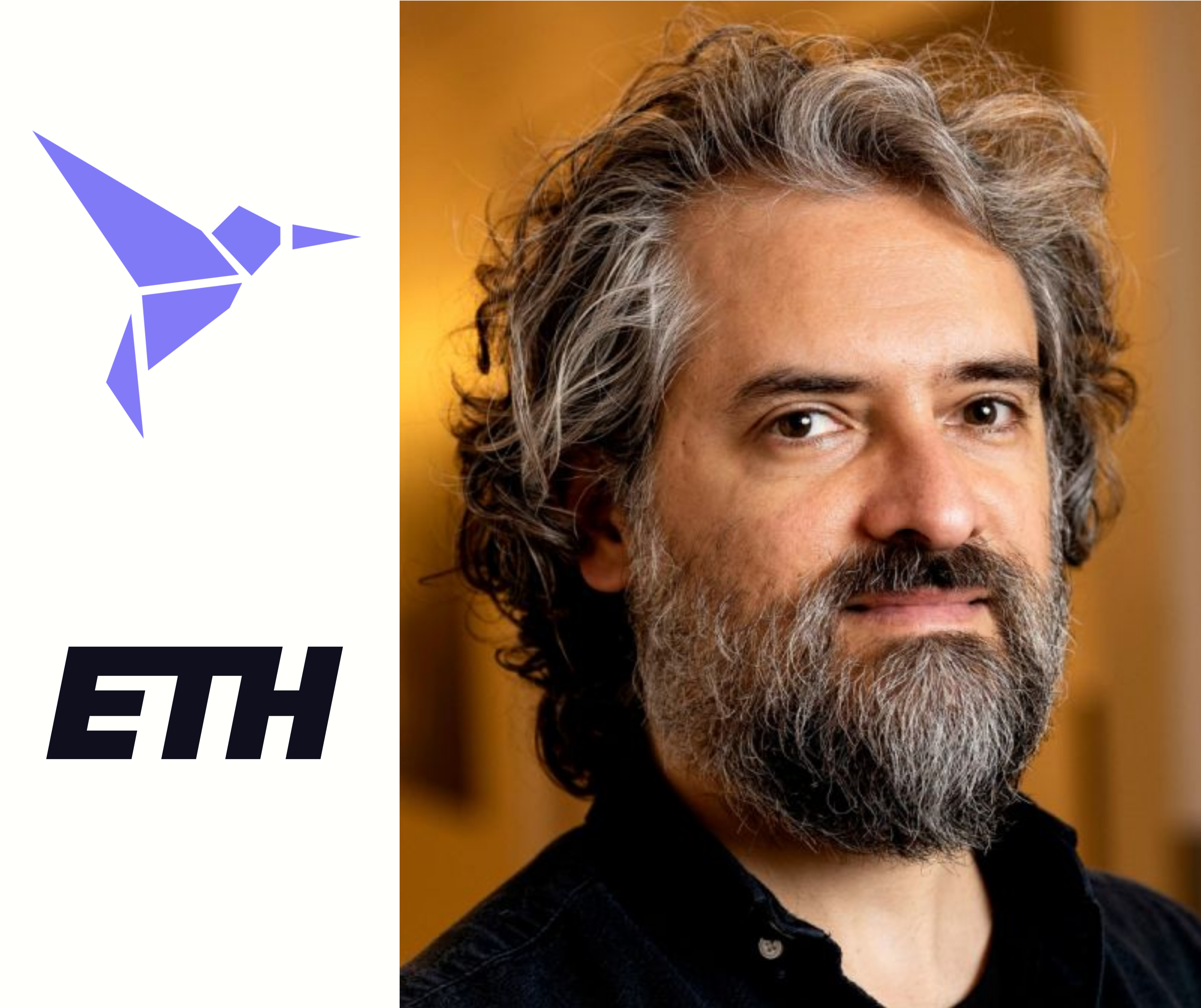
Home Publications Investors News Get Started IFLAI’s CTO Leads Advanced Machine Learning Course at ETH Zurich In recognition of his expertise, Giovanni Volpe, CTO of IFLAI, was invited to conduct a course on “Advanced Machine Learning Techniques Applied to Microscopy Data Analysis” at ETH Zurich. The session began on October 5, 2022. Fifteen doctoral students and post-docs participated in the event, where they delved into topics such as Dense Neural Networks, Recurrent Neural Networks, Transformers, and more. The session highlighted the practical impact of IFLAI’s technological prowess on the academic and research communities. This collaboration showcases IFLAI’s commitment to fostering educational advancements in machine learning and reinforces IFLAI’s position as a leader in technological advancements. Read more Recent Posts September 18, 2023 Giovanni Volpe awarded the Faculty of Science’s 2023 Research Award April 5, 2023 IFLAI participates in GAIA Conference 2024, sharing the stage with global AI leaders February 20, 2023 Giovanni Volpe Shares Machine Learning Expertise at ICFO January 24, 2023 IFLAI has been awarded the prestigious ERC Proof of Concept Grant October 7, 2022 Kommentar på Clines Scientifics inledda samarbete med IFLAI gällande utveckling av AI-mjukvara för CellRace-projektet Publications Investors News Contact Us AddressHasselvägen 85, Mölnlycke Emailcontact@iflai.com Get Connected
Microplankton life histories revealed by holographic microscopy and deep learning
Home Investors News Publications Get Started Microplankton life histories revealed by holographic microscopy and deep learning Harshith Bachimanchi, Benjamin Midtvedt, Daniel Midtvedt, Erik Selander & Giovanni Volpe eLife (2022) Abstract The marine microbial food web plays a central role in the global carbon cycle. However, our mechanistic understanding of the ocean is biased toward its larger constituents, while rates and biomass fluxes in the microbial food web are mainly inferred from indirect measurements and ensemble averages. Yet, resolution at the level of the individual microplankton is required to advance our understanding of the microbial food web. Here, we demonstrate that, by combining holographic microscopy with deep learning, we can follow microplanktons throughout their lifespan, continuously measuring their three-dimensional position and dry mass. The deep-learning algorithms circumvent the computationally intensive processing of holographic data and allow rapid measurements over extended time periods. This permits us to reliably estimate growth rates, both in terms of dry mass increase and cell divisions, as well as to measure trophic interactions between species such as predation events. The individual resolution provides information about selectivity, individual feeding rates, and handling times for individual microplanktons. The method is particularly useful to detail the rates and routes of organic matter transfer in micro-zooplankton, the most important and least known group of primary consumers in the oceans. Studying individual interactions in idealized small systems provides insights that help us understand microbial food webs and ultimately larger-scale processes. We exemplify this by detailed descriptions of micro-zooplankton feeding events, cell divisions, and long-term monitoring of single cells from division to division. View publication
Label-free nanofluidic scattering microscopy of size and mass of single diffusing molecules and nanoparticles
Home Publications Investors News Get Started Label-free nanofluidic scattering microscopy of size and mass of single diffusing molecules and nanoparticles Barbora Špačková, Henrik Klein Moberg, Joachim Fritzsche, Johan Tenghamn, Gustaf Sjösten, Hana Šípová-Jungová, David Albinsson, Quentin Lubart, Daniel van Leeuwen, Fredrik Westerlund, Daniel Midtvedt, Elin K. Esbjörner, Mikael Käll, Giovanni Volpe & Christoph Langhammer Nature Methods (2022) Abstract Label-free characterization of single biomolecules aims to complement fluorescence microscopy in situations where labeling compromises data interpretation, is technically challenging or even impossible. However, existing methods require the investigated species to bind to a surface to be visible, thereby leaving a large fraction of analytes undetected. Here, we present nanofluidic scattering microscopy (NSM), which overcomes these limitations by enabling label-free, real-time imaging of single biomolecules diffusing inside a nanofluidic channel. NSM facilitates accurate determination of molecular weight from the measured optical contrast and of the hydrodynamic radius from the measured diffusivity, from which information about the conformational state can be inferred. Furthermore, we demonstrate its applicability to the analysis of a complex biofluid, using conditioned cell culture medium containing extracellular vesicles as an example. We foresee the application of NSM to monitor conformational changes, aggregation and interactions of single biomolecules, and to analyze single-cell secretomes. View publication Publications Investors News Contact Us AddressHasselvägen 85, Mölnlycke Emailcontact@iflai.com Get Connected
IFLAI wins the pitching competition at the Startup Camp 2022
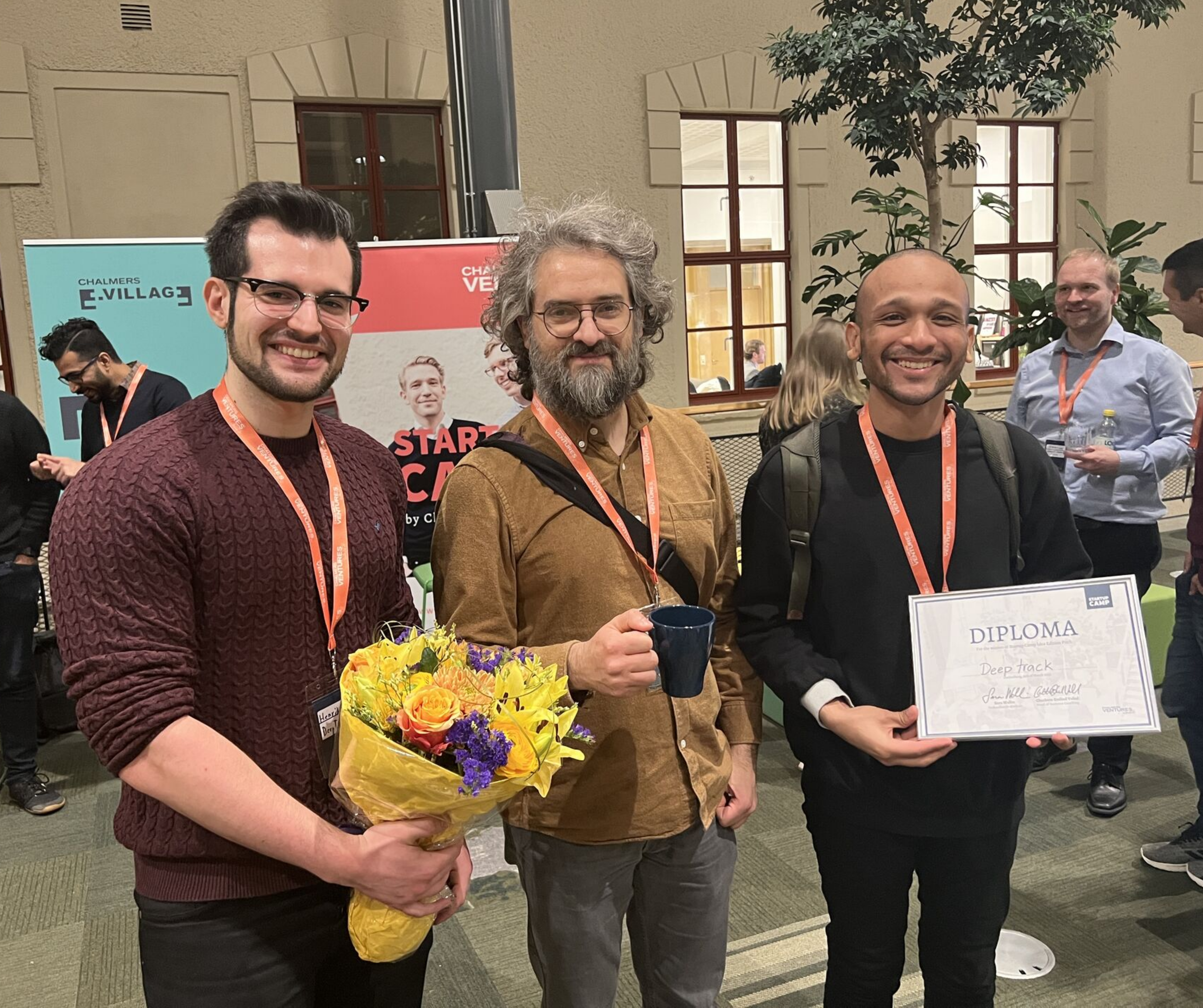
Home Publications Investors News Get Started IFLAI wins the Startup Camp 2022 Pitching Competition Picture by Jonas Sandwall, Chalmers Ventures. IFLAI emerged victorious at the Startup Camp 2022 pitching competition, organized by Chalmers Ventures. The competition, which took place on Tuesday, 15 March 2022, from 16:00 to 19:00, featured ten ambitious teams that had undergone rigorous training at the Startup Camp, where they developed and honed their company ideas. These ten teams took to the stage to pitch their innovative business concepts to a discerning panel of entrepreneurial experts, their fellow competitors, and all business coaches at Chalmers Ventures. In a tightly contested competition, IFLAI distinguished itself by securing first place, demonstrating exceptional creativity, business acumen, and the potential for significant impact in their industry. The victory at the Startup Camp 2022 pitching event not only highlights IFLAI’s promising future but also underscores the importance of platforms like Chalmers Ventures in nurturing and recognizing emerging talent in the entrepreneurial ecosystem. Read more Recent Posts November 25, 2023 IFLAI’s Chief AI Engineer spearheads AI dialogue at Future Impact Summit 2023 September 18, 2023 Giovanni Volpe awarded the Faculty of Science’s 2023 Research Award April 5, 2023 IFLAI participates in GAIA Conference 2024, sharing the stage with global AI leaders February 20, 2023 Giovanni Volpe Shares Machine Learning Expertise at ICFO January 24, 2023 IFLAI has been awarded the prestigious ERC Proof of Concept Grant Publications Investors News Contact Us AddressHasselvägen 85, Mölnlycke Emailcontact@iflai.com Get Connected
Extracting quantitative biological information from bright-field cell images using deep learning
Home Publications Investors News Get Started Extracting quantitative biological information from bright-field cell images using deep learning Saga Helgadottir, Benjamin Midtvedt, Jesus Pineda, Alan Sabirsh, Caroline B. Adiels, Stefano Romeo, Daniel Midtvedt, & Giovanni Volpe. Biophysics Reviews (2021) Abstract Quantitative analysis of cell structures is essential for biomedical and pharmaceutical research. The standard imaging approach relies on fluorescence microscopy, where cell structures of interest are labeled by chemical staining techniques. However, these techniques are often invasive and sometimes even toxic to the cells, in addition to being time consuming, labor intensive, and expensive. Here, we introduce an alternative deep-learning–powered approach based on the analysis of bright-field images by a conditional generative adversarial neural network (cGAN). We show that this is a robust and fast-converging approach to generate virtually stained images from the bright-field images and, in subsequent downstream analyses, to quantify the properties of cell structures. Specifically, we train a cGAN to virtually stain lipid droplets, cytoplasm, and nuclei using bright-field images of human stem-cell–derived fat cells (adipocytes), which are of particular interest for nanomedicine and vaccine development. Subsequently, we use these virtually stained images to extract quantitative measures about these cell structures. Generating virtually stained fluorescence images is less invasive, less expensive, and more reproducible than standard chemical staining; furthermore, it frees up the fluorescence microscopy channels for other analytical probes, thus increasing the amount of information that can be extracted from each cell. To make this deep-learning–powered approach readily available for other users, we provide a Python software package, which can be easily personalized and optimized for specific virtual-staining and cell-profiling applications. View publication Publications Investors News Contact Us AddressHasselvägen 85, Mölnlycke Emailcontact@iflai.com Get Connected
Quantitative digital microscopy with deep learning
Home Publications Investors News Get Started Quantitative digital microscopy with deep learning Benjamin Midtvedt, Saga Helgadottir, Aykut Argun, Jesus Pineda, Daniel Midtvedt, and Giovanni Volpe. Applied Physics Reviews (2021) Abstract Video microscopy has a long history of providing insight and breakthroughs for a broad range of disciplines, from physics to biology. Image analysis to extract quantitative information from video microscopy data has traditionally relied on algorithmic approaches, which are often difficult to implement, time-consuming, and computationally expensive. Recently, alternative data-driven approaches using deep learning have greatly improved quantitative digital microscopy, potentially offering automatized, accurate, and fast image analysis. However, the combination of deep learning and video microscopy remains underutilized primarily due to the steep learning curve involved in developing custom deep-learning solutions. To overcome this issue, we introduce software, DeepTrack 2.0, to design, train, and validate deep-learning solutions for digital microscopy. We use this software to exemplify how deep learning can be employed for a broad range of applications, from particle localization, tracking, and characterization, to cell counting and classification. Thanks to its user-friendly graphical interface, DeepTrack 2.0 can be easily customized for user-specific applications, and thanks to its open-source, object-oriented programing, it can be easily expanded to add features and functionalities, potentially introducing deep-learning-enhanced video microscopy to a far wider audience. View publication Publications Investors News Contact Us AddressHasselvägen 85, Mölnlycke Emailcontact@iflai.com Get Connected